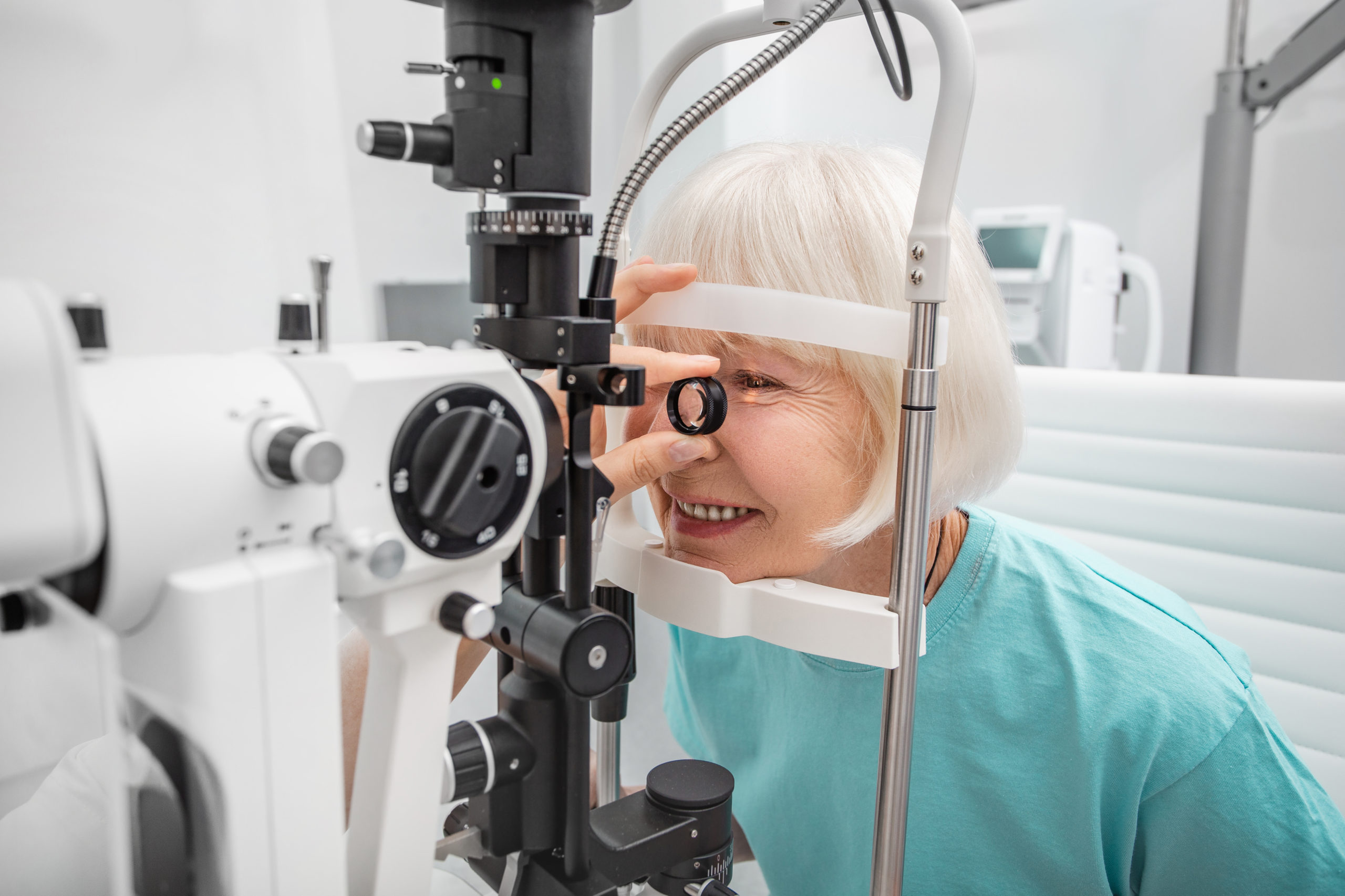
One of the many common tests performed at Tower Clock Eye Center is the slit lamp exam. This exam is done with a microscope and bright light that gives your eye doctor the ability to examine the many structures around and inside the eye. The slit lamp exam is a vital exam to determine the overall health of the eye and helps detect eye disease.
Preparing for the exam:
The eyes need no special prep to be examined with a slit lamp, however the eyes are sometimes dilated (the pupil is widened to allow for a better view of the back of the eye) using eye drops. Eyes are usually blurry once dilated and they’ll be sensitive to light. Patients are advised to arrange for a drive home following a dilated exam. Sunglasses are also recommended.
The slit lamp exam:
The slit lamp microscope will swing around in front of the exam seat and patients will place his or her chin on the rest, with the forehead against a rounded band. This is a comfortable position and allows the entire head to remain steady as the doctor peers into the patient’s eye. The doctor will sit in front of you and use the scope to examine several parts of the eye (see below). During the exam the lamp will be turned on and a thin, narrow light will help the doctor look into the eye. The light will be bright, but it won’t cause damage. In addition to the dilating drops, some patients will be given a drop with a specific dye in it to better exam the front structures of the eye.
What is examined:
Cornea: The cornea is the clear, curved front part of the eye. The slit lamp test can help diagnose dry eye by examining this part of the eye. The thickness (and thinness) of the cornea can be determined which can show several corneal-specific abnormalities causing blurred or loss of vision.
Lens: The clear portion behind the pupil that focuses light is the lens. Typically the lens is examined for the presence of a cataract, the clouding of the body’s natural lens. As we all know, cataracts happen as we age and are easily fixed with cataract surgery.
Sclera: The sclera is the white part of the eye and consists of strong fibrous tissue that protects the outermost layers of the eye. Slit lamp exams help doctors see inflammation, discoloration and other abnormalities of the sclera. The conjunctiva, an extremely thin, transparent layer over the sclera, can also be examined for conjunctivitis, commonly known as pink eye, as well as other diseases.
Retina: The retina is the thin layer of photoreceptor cells at the back of the eye. These cells send signals to the optic nerve which relays light and vision to the brain. Slit lamp exams can determine many conditions including age-related macular degeneration and retinitis pigmentosa. Another issue to be examined for are torn or detached retinas – a critical issue that requires immediate treatment to prevent blindness.
Optic nerve: As mentioned above, the optic nerve attaches to the brain. This nerve can be damaged by glaucoma, which is heightened pressure within the eye. Glaucoma works slowly and can be difficult to diagnose but a slit lamp exam can help diagnose it early, leading to treatment and prevents loss of vision.
Lid and lashes: The structures around the eyeball are examined for signs of inflammation, infection or any disease. Bumps on eyelids are often evaluated with a slit lamp.
Cataract surgery is one of the most common and successful procedures performed worldwide, restoring vision for millions of individuals each year. Tower Clock Eye Center surgeons offer the latest technology and intraocular lens (IOL) options for cataract surgery to assure the best possible outcomes. While the focus is often on the removal of the clouded... read more
All Tower Clock Eye Center offices will be closed on Thursday, July 4, to celebrate the holiday. We'll reopen on Friday with our normal schedule. We wish you all a happy and safe holiday!